Tuesday, November 10, 2015

A 60 years old woman with gradual paralysis and severe back pain for the last few weeks

This is a HIPAA de-identified open-online-patient-record, posted on autumn 2015 after collecting informed patient consent (form downloadable here ) by LNMCH research assistant and patient-information-communication-executive and discussion initiated by patient's primary care physician in-charge: 

Conversational clinical decision support from online learning forum:

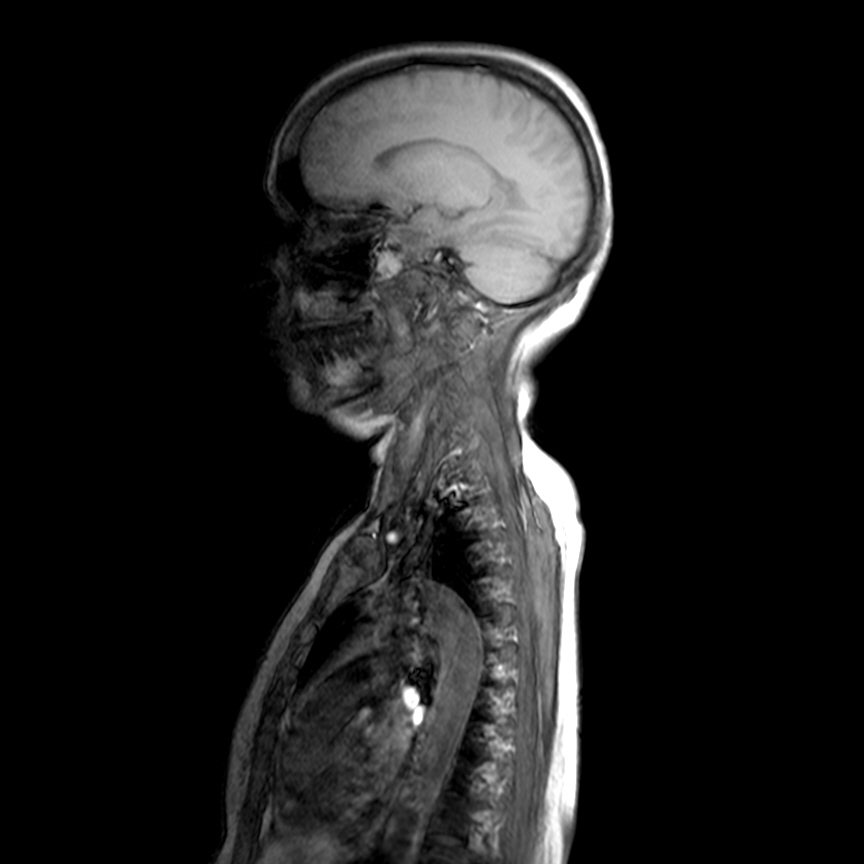
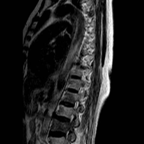
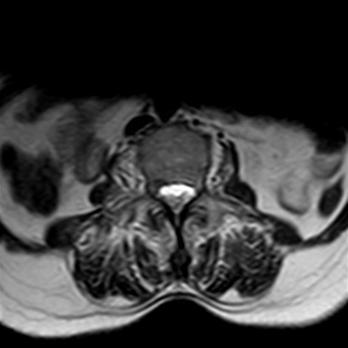
Needed help for this currently admitted 60 years old woman with gradual paralysis and severe back pain for the last few weeks. On examination there was slight gibbus and absent lower limb reflexes and MRI shows a lesion at L3 as well as T3.

Online forum conversations:


Santosh Sapkota Tuberculosis?
Abhimanyu Jha NHL/plasma cell neoplasm??
Rakesh Biswas Thanks Dr Santosh and Dr Abhimanyu for these inputs. It shows that we may need a biopsy to microscopically differentiate between these possibilities. Another thought that has been with me for a few days now is how we are ruled by our quest to define both the macro-pathology (reflected in our patient's clinical and radiological images) and micro-pathology (which currently invariably requires an invasive biopsy toward microscopy). Can technology someday help us to visualize microscopic images on MRIs (or something similar)?
Santosh Sapkota That's money right there $$$$$$
Rakesh Biswas
Write a reply...
Nitin Garg This looks like metastasis in view of multiple discontinuous vertebral body involvement, and also posterior elements involved. Usg guided FNAC/ biopsy can be done at L2 level.
There are high strength MRI under development to give microscopic picture but these are still not feasible for clinical use.

Rakesh Biswas Thanks Dr Nitin for these valuable comments. We shall try to get her FNAC done from the posterior elements tomorrow (Dr Deepika, MrKuldeep). Vipul would be good to have your inputs on image processing technology in radio-diagnosis progressing toward microscopy.
Nitin Garg Do we have any work up done for a primary lesion?
Rakesh Biswas We saw something suspicious in the CXR pa view in the left lower zone although the left lateral view didn't support it. We shall get an HRCT today (Dr DeepikaJemmima we would need to take a re-look at her breasts for a primary there. Can also ask Dr Swagata and Dr Bharati to evaluate for that).
Swagata Brahmachari Ya we can examine the pt in sopd tommorrow
Rakesh Biswas Abhimanyu you remember how you had FNACed a similar patient on the basis of CXR pa alone and given us the diagnosis. I wish we had an Abhimanyu in our pathology department here. smile emoticon
Abhimanyu Jha Thank you sir for the complement

Below are the images of doctor notes and patient radiology and investigations uploaded in autumn 2015 by LNMCH research assistant and patient-information-communication-executive: 

Doctor's notes:


Investigation images:


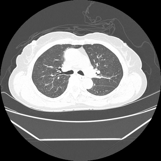
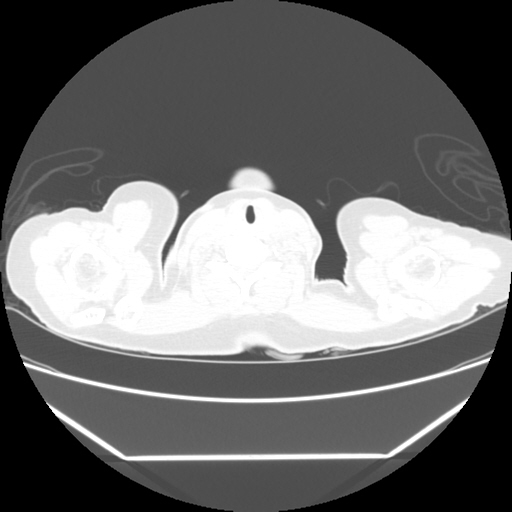
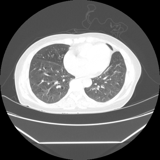
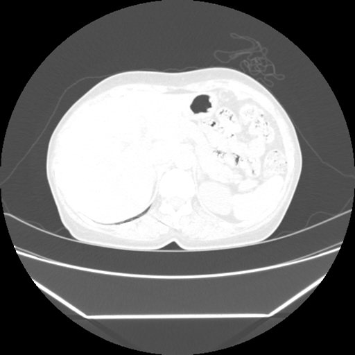
Radiological images:


HRCT is also done on 13/11/15


USG report & images:


No comments:

Post a Comment